This page is generated from the same slides.md source as the Slidev deck.
Its sections flow as a regular document, while the source-estimate viewer and
sensor demonstrations remain interactive.
Building Reproducible Neuroscience with NeuroWaves
Core Technology Platforms, Knowledge Exchange Series
BioMedical Imaging Core · NYUAD Research Institute
Hadi Zaatiti · April 2026
Section 2
Today's Roadmap
1
The NeuroWaves Lab Who we are, What do we do ?
2
Two MEG technologies at NYUAD SQUID-based KIT system and OPM-based HEDscan system
3
Projects and collaborations From Electrical Engineering, to Neuroscience, Psychology and soon... Clinical Applications
4
How We Work Collaborative platforms, Community-driven documentation, Reproducible pipelines
5
Next steps
Section 3
The NeuroWaves Lab
NeuroWaves is a core facility within NYUAD's BioMedical Imaging Core, part of the Core Technology Platforms.
We provide training, experiment code, processing pipelines, and comprehensive documentation for researchers using Magnetoencephalography (MEG) and Electroencephalography (EEG) at NYUAD.
The Team
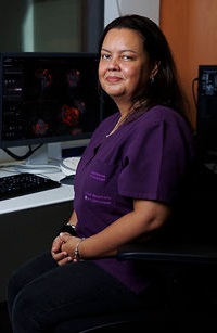
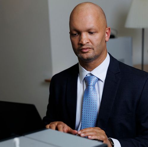
Hadi Zaatiti
MEG Research Scientist
Haidee Paterson
MRI Instrumentation Specialist
Osama Abdullah
MRI Physicist CTP Director
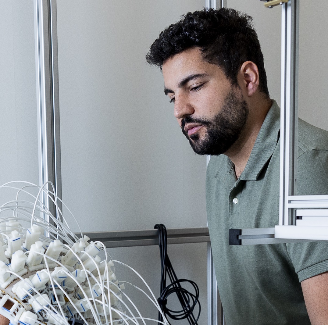
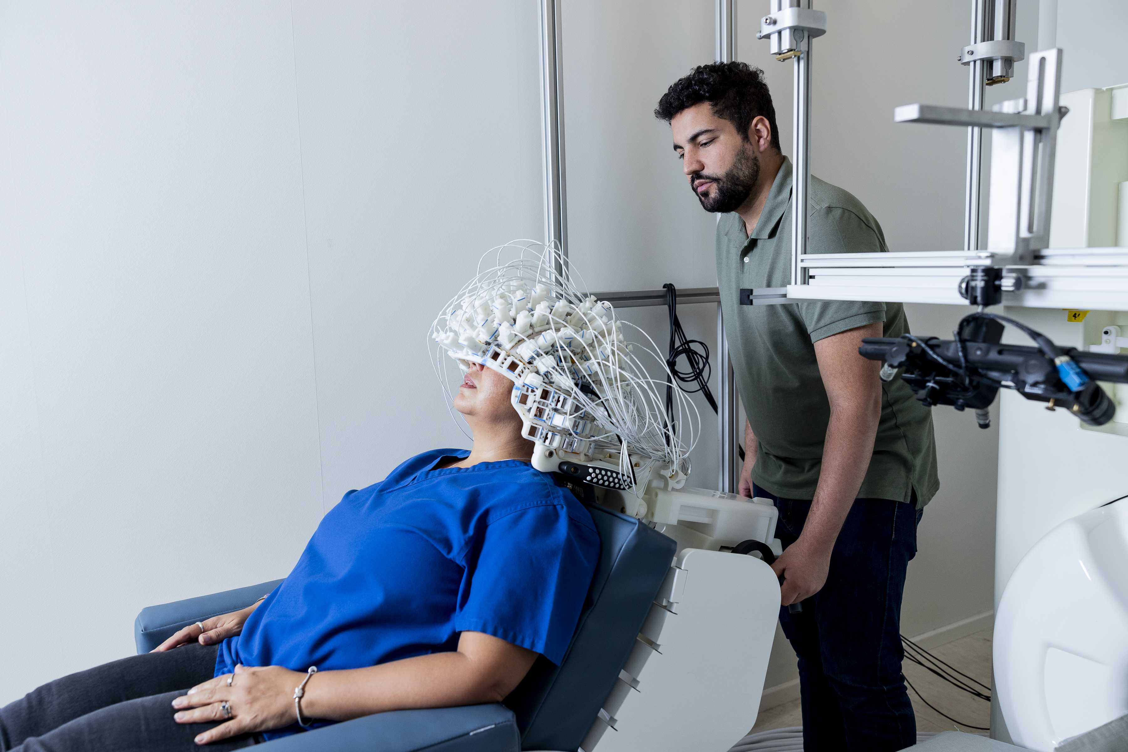
OPM-MEG recording session at the NeuroWaves lab
Section 4
The NeuroWaves Laboratory
What is MEG/EEG?
Magnetoencephalography (MEG) and Electroencephalography (EEG) non-invasively measure:
the tiny magnetic femtoteslas (fT, 10⁻¹⁵ T)/electric microvolts (μV, 10⁻⁶ V) fields produced by neuronal activity. Sensors with extreme sensitivity are required.
Spatio-Temporal Resolution
Low Temporal → High Temporal
Low Spatial → High Spatial
fMRI
PET
MEG
EEG
fNIRS
~ 1 ms
MEG and EEG Temporal Resolution
~2-3 mm
MEG Spatial Resolution
100%
Non-Invasive
2
MEG Systems at NYUAD
Section 5
Two MEG technologies at NYUAD
How MEG Sensors Work
Two technologies measure the brain's magnetic fields.
SQUID Operation · 1: The Brain, where the signal originates
Loading interactive figure…
A localized cortical region activates. MEG signals primarily originate from sulci, where current flows tangentially to the scalp, producing an external magnetic field detectable outside the head.
Section 7
Two MEG technologies at NYUAD
SQUID Operation · 2: Neurons, how the field is generated
Loading interactive figure…
A single neuron produces only a fraction of a femtotesla, far below detector noise. The resulting ~100 fT field is roughly 500 million times weaker than Earth's magnetic field, which is why MEG requires heavy magnetic shielding.
Section 8
Two MEG technologies at NYUAD
SQUID Operation · 3: The Sensor, superconducting pickup at 4 K
Loading interactive figure…
Two coils wound in opposition, connected in series, form an axial gradiometer. The closed superconducting loop threads a SQUID ring, whose two Josephson junctions convert the flux difference between the coils into voltage. The assembly sits in a liquid helium dewar at 4 K, about 20 mm from the scalp.
Section 9
Two MEG technologies at NYUAD
SQUID Operation · 4: The Signal, what we record
Loading interactive figure…
MEG signal digitized at 2 kHz, noise floor 4 fT/√Hz. The classic M100/M200 evoked response emerges after averaging. Ultra-low noise, fixed sensor position.
Section 10
Two MEG technologies at NYUAD
OPM Operation · 1: The Brain, same neural source
Loading interactive figure…
Same physics, same source. Cortex activates, fields emerge from sulci. What changes is how we sense them.
Section 11
Two MEG technologies at NYUAD
OPM Operation · 2: Neurons, same pyramidal source
Loading interactive figure…
Synchronous postsynaptic currents, ~100 fT magnetic field, right-hand rule. The signal is identical. OPM sensors just sit closer to it.
Section 12
Two MEG technologies at NYUAD
OPM Operation · 3: The Sensor, atomic magnetometer at room temp
Loading interactive figure…
A 795 nm laser aligns Rb-87 electron spins inside a vapor cell. The brain's field causes the spins to precess, which changes light absorption. A photodetector reads the change, all at room temperature, directly on the scalp.
Section 13
Two MEG technologies at NYUAD
OPM Operation · 4: The Signal, stronger but noisier
The Magnetically Shielded Room (MSR) built from multiple layers of mu-metal (high-permeability Ni-Fe alloy) and aluminum. Together these attenuate external magnetic noise by roughly six orders of magnitude, essential because the brain's fields (~100 fT) are a 100 million times weaker than Earth's field. Today, both SQUID-MEG and OPM-MEG systems operate inside.
Location
NYUAD · A2-008 BioMedical Imaging Core
Shielding
Mu-metal + aluminum multi-layer construction
Noise reduction
~10⁶× attenuation DC to kHz range
Section 15
Two MEG technologies at NYUAD
SQUID-MEG: The KIT System
Eagle Technology / Kanazawa Institute of Technology
208 SQUID Sensors
First-order axial gradiometers for measuring brain magnetic fields
16 Magnetometers
SQUID magnetometers for environmental noise suppression
Cryogenic Cooling
Liquid helium dewar at ~4 K maintaining superconductivity
2 kHz Sampling
High-fidelity continuous data acquisition
4 fT/√Hz Noise Floor
Ultra-low noise at 180 Hz
12 years old system
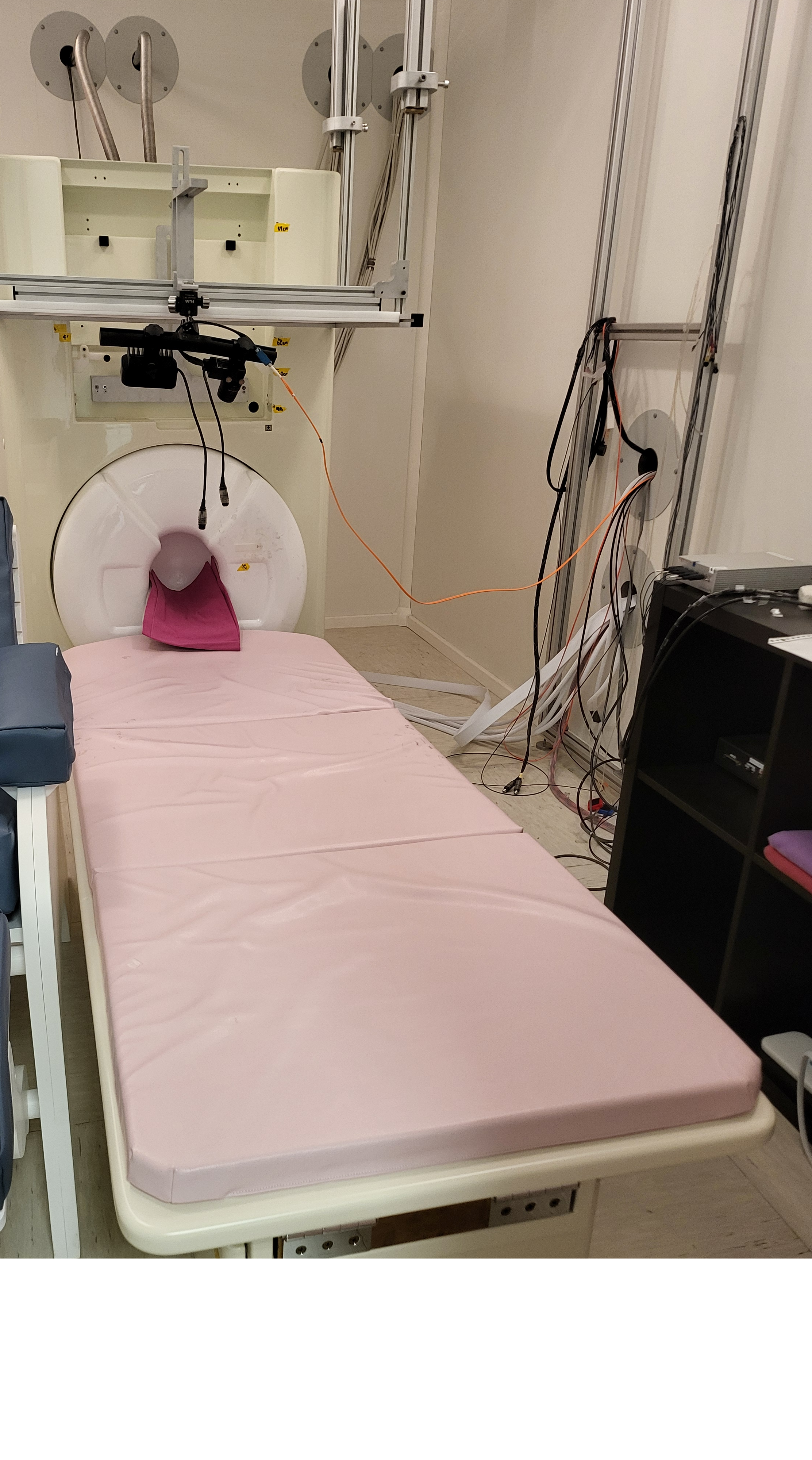
KIT 208-channel SQUID-MEG system
Section 16
Two MEG technologies at NYUAD
OPM-MEG: The HEDscan System
FieldLine Medical Inc., Boulder, CO, USA
96 OPM Sensors
Optically pumped magnetometers, no cryogenic cooling required
Flexible Helmet
Wearable sensor holder adaptable to individual head shapes
On-Scalp Recording
Sensors positioned directly on scalp for higher signal amplitude
5 kHz Sampling
High-frequency acquisition for detailed signal capture
Room Temperature
No liquid helium, lower operational costs and complexity
2 years old
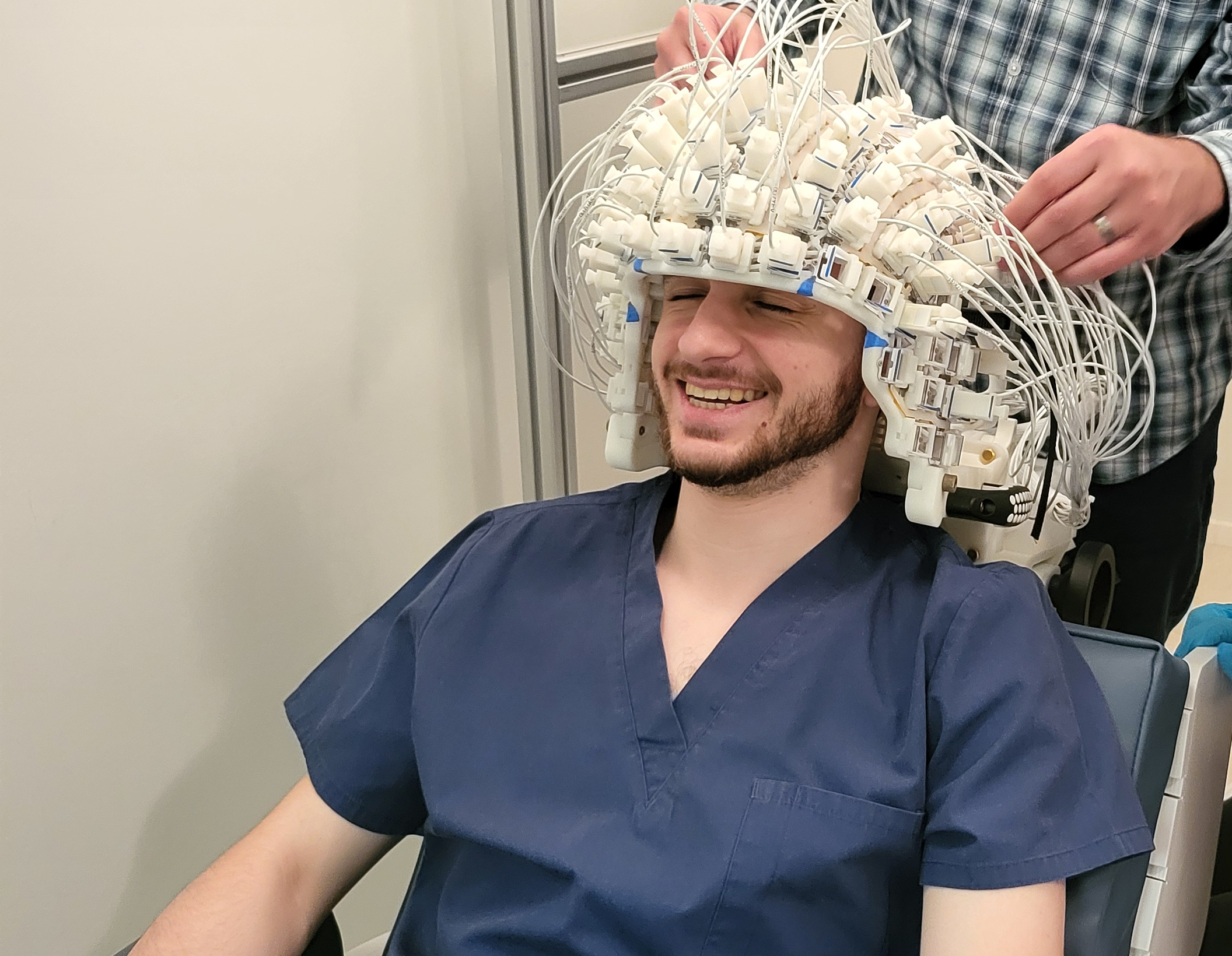
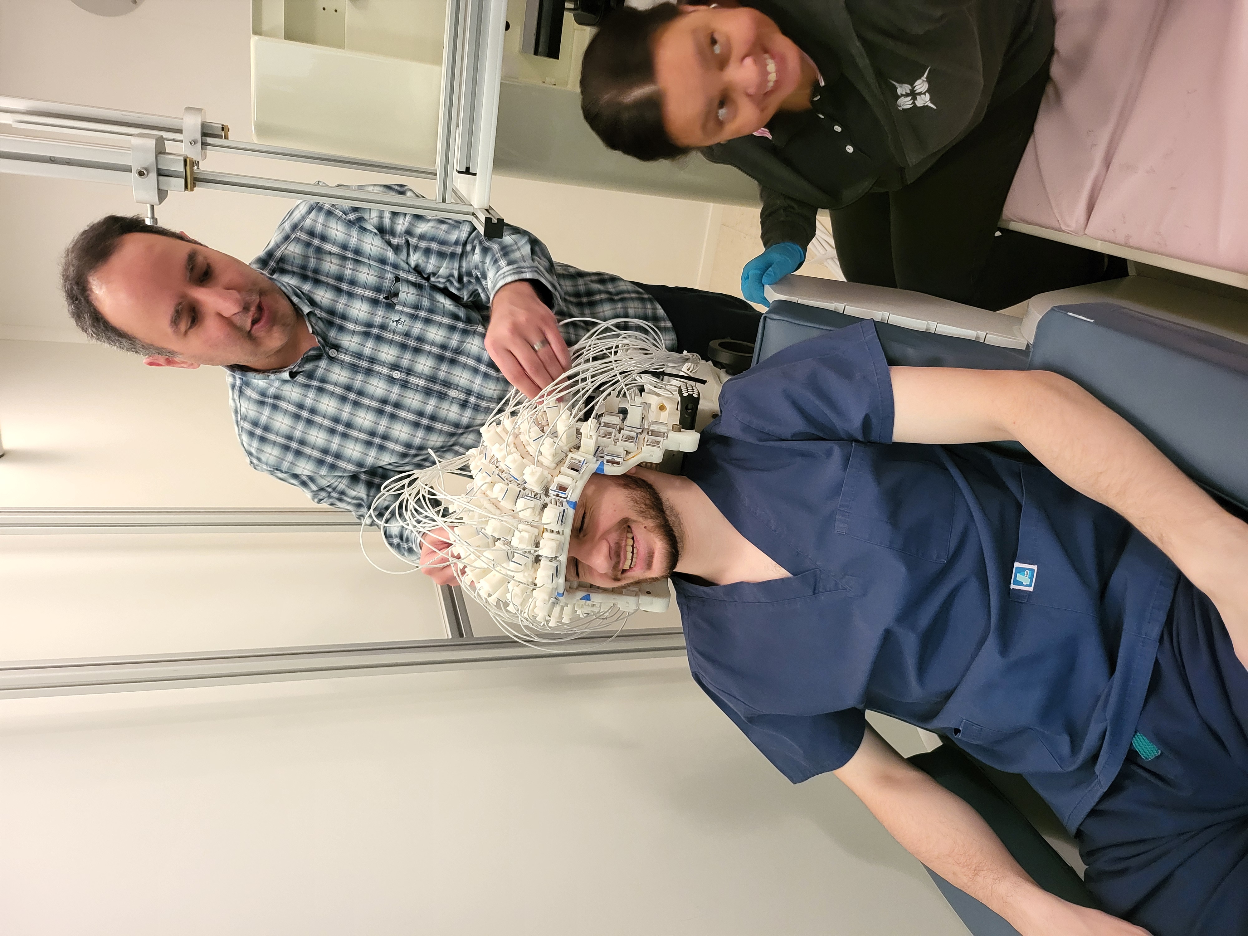
FieldLine HEDscan OPM-MEG helmet
Section 17
Two MEG technologies at NYUAD
Data processing: preprocessing, time and frequency analysis
Preprocessing: band pass filter 0.1 to 200 Hz with a 50 Hz notch, bad channel interpolation from spatial neighbours, ICA removes ECG, EOG and eyeblinks, HPI tracking corrects head motion, then epoching around stimulus events.
Evoked Response Field (ERF)
Averaging trials time locked to a stimulus reveals M100, M200, and later components. Captures phase locked activity only.
Frequency domain: α example (time then spectrum)
A 10 Hz α sinusoid (top) becomes a sharp peak inside the α band (bottom).
δ Delta 1 to 4 Hz deep sleep, unconscious processes
θ Theta 4 to 8 Hz drowsiness, memory encoding, meditation
β Beta 13 to 30 Hz active thinking, motor control, focus
γ Gamma 30 to 80 Hz perception, attention, neural binding
Section 18
Two MEG technologies at NYUAD
Data processing: source localization
We record magnetic fields at 208 or 96 sensors. From those recordings, what brain activity produced them?
An analogy Imagine you only see the shadow of an object on the ground. Different objects can cast very similar shadows. From the shadow alone, many objects fit.
Source localization The sensor data is the shadow. The cortical activity is the object. We use a head model from the MRI and a few mild assumptions to pick the most plausible source pattern.
Dipole fitting focal sources
MNE, sLORETA distributed cortex
Beamforming LCMV, DICS
Section 19
Two MEG technologies at NYUAD
Source estimates: Visual and Auditory
Drag each slider to scrub through time. Left: visual (checkerboard). Right: auditory (tone).
Loading interactive figure…
Section 20
Projects and Collaborations
Current research portfolio on the MEG platform
16 active or planned projects · 4 principal investigators · 11 current user researchers · spanning Arabic (Emirati and Egyptian), English, Mandarin, Korean, Italian, and bilingual populations.
Cognitive Neuroscience6 projects
Visual Working Memory Hamkari · PI Melcher · N=20 · planned
Visual Imagery Zhang · PI Melcher · N=20 · in progress
Object Categorization PI Melcher · N=20 · planned
HEAL Project Al Shaali, Enriquez · PI Melcher · N=35 · in progress
Visual Crowd Perception Al Shaali, Ezzedine · PI Melcher · N=20 · completed
Working Memory Satheesh · PI Sreenivasan · N=20 · in progress
Psycholinguistics10 projects · 5 languages
SPROUSE LAB
Arabic (Emirati) Elmallah · N=30 · in progress
Arabic (Egyptian) Elmallah · N=30 · in progress
Mandarin Elmallah · N=30 · in progress
Korean Park · N=30 · planned
Italian Cerrone · N=30 · planned
ALMEIDA LAB
Arabic and English bilingual N=36 to 48 · planned
Arabic language N=36 to 48 · planned
English language N=TBD · planned
Masked Priming (English) Alobaidalla · planned
Masked Priming (English) Jemi · planned
Section 21
Projects and Collaborations
SQUID vs OPM comparison project
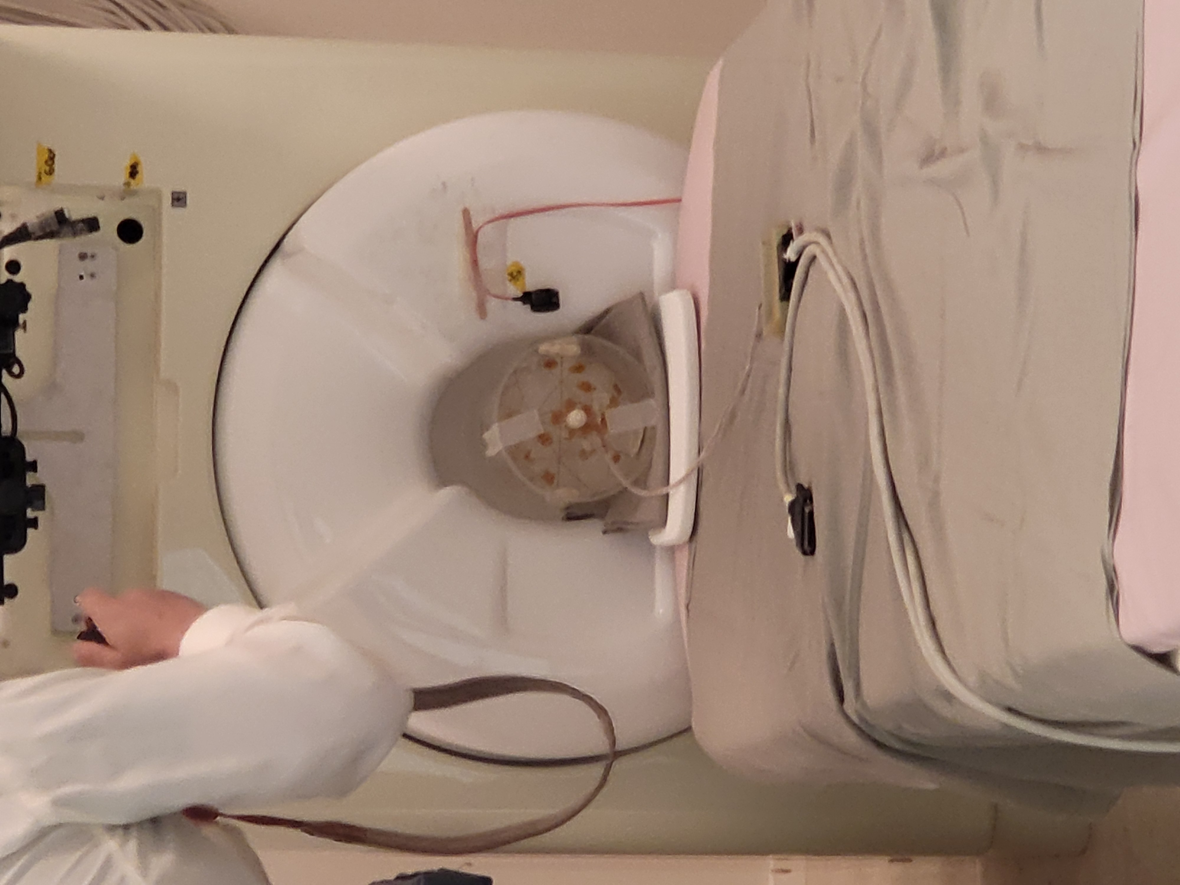
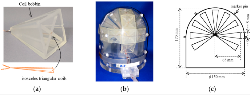
Photograph and configuration of the dry type MEG phantom: (a) closeup view of the triangular coil; (b) photograph of the dry type MEG phantom; (c) schematic and dimensions of the phantom.
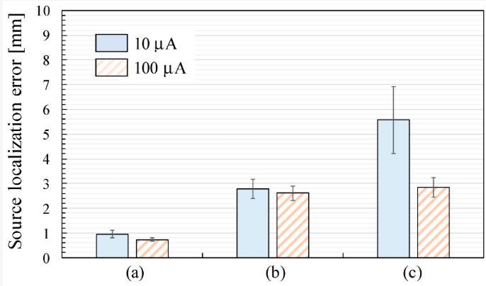
Source localization error defined as the displacement between the effectual and estimated ECD positions: (a) SQUID MEG system; (b) OPM MEG system (closest position); (c) OPM MEG system (lift off position).
Oyama, D. and Zaatiti, H. (2025) Phantom Based Approach for Comparing Conventional and Optically Pumped Magnetometer MEG Systems Sensors, 25(7), 2063
SQUID vs OPM accuracy 1.07 ± 0.17 mm with 202 SQUID sensors vs 3.48 ± 0.58 mm with 90 OPM sensors at closest position
Signal strength OPM detected 3 to 4 times higher amplitude thanks to on scalp proximity
40 vs 90 sensors A 40 channel SQUID subset reached accuracy comparable to 90 channel OPM
Section 22
Projects and Collaborations
OPM calibration project
Condition
GOF (mean ± SD)
ORG
SPH
Close / 0.01 mA
99.22 ± 0.40%
99.90 ± 0.05%
Close / 0.1 mA
99.23 ± 0.40%
99.92 ± 0.04%
Far / 0.01 mA
99.21 ± 0.25%
99.75 ± 0.12%
Far / 0.1 mA
99.32 ± 0.24%
99.88 ± 0.08%
Goodness of fit of equivalent current dipole estimates. ORG is the sensor localization function originally implemented in the OPM smart helmet; SPH is the spherical coil array calibration. Close and Far refer to sensor placement on the phantom dome; 0.01 and 0.1 mA are the dipole currents. SPH improves GOF across all four conditions.
Adachi, Y., Oyama, D., Uehara, G., and Zaatiti, H. (2025) Sensor Array Geometry Acquisition by Spherical Coil Array for OPM Based MEG System IEEE Sensors Journal, 25(22), 41200 to 41208
Calibration method A 150 mm spherical array of 16 circular coils replaces the built in smart helmet localization and recovers each OPM position, orientation, and sensitivity from a single calibration recording.
Two modes, two currents Close (sensor tips in contact with the phantom dome) and Far (shifted by about 20 mm). Dipole currents of 0.01 and 0.1 mA. 96 OPM sensors in close, 95 in far. 49 ECDs on a dry type phantom.
Main result SPH matches ORG positions within 1 mm but recovers sensor orientations on average 7.2° away from factory values. ECD localization variability drops by about 50% in close mode, confirmed by 95% Bayesian credible intervals.
Section 23
Projects and Collaborations
MEG Demo Classes
Hands on lab sessions for NYUAD classes
Cognitive Neuroscience Resting state and attention paradigm with live source localization demo
Biopsychology Resting state and auditory vs visual vs motor experiment with real time analysis
Bioengineering Full KIT and OPM system tour, resting state and multi sensory experiment
What Students Experience
Lab tour
Participant preparation and laser scan
Live SQUID sensitivity demo
Real experiments
On the spot data analysis and discussion
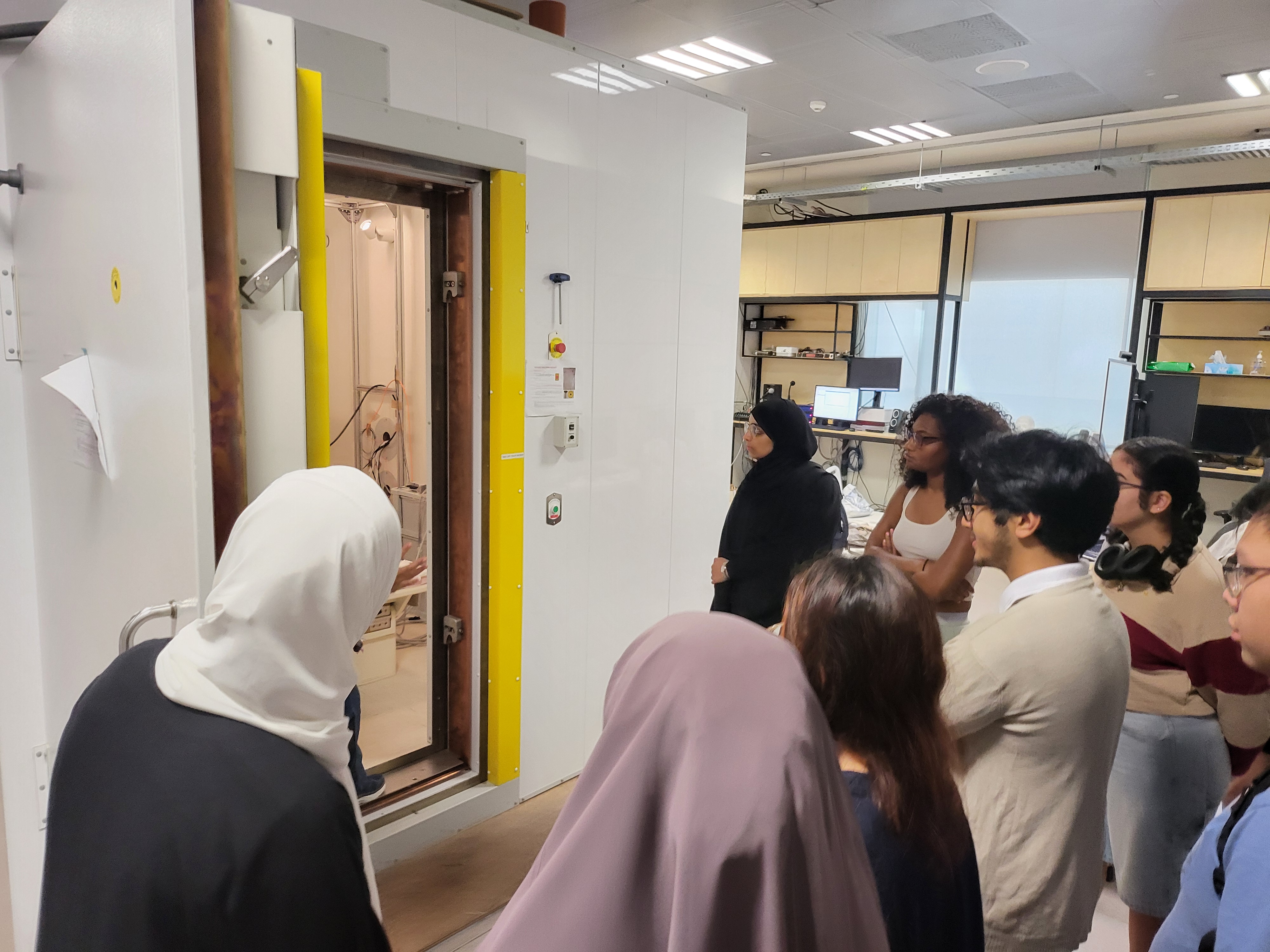
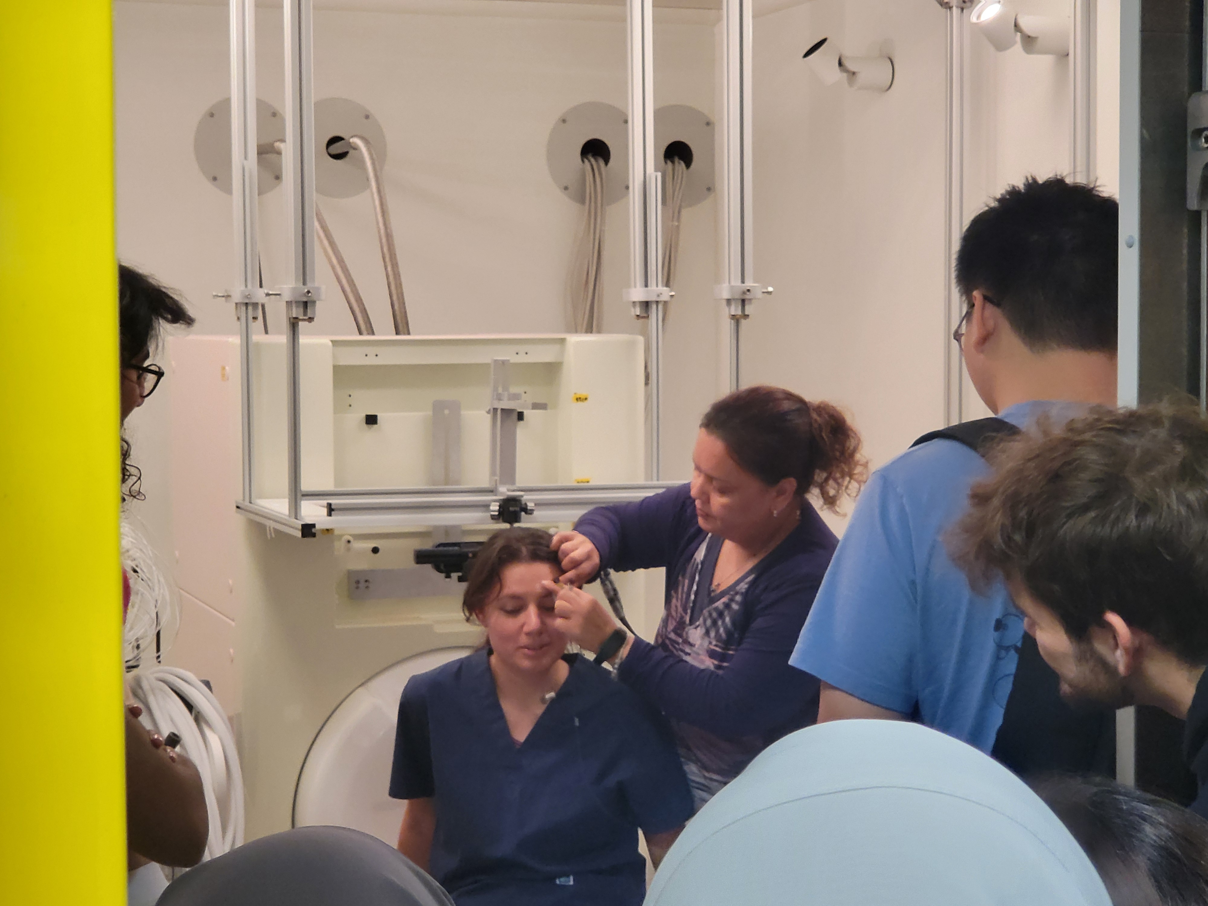
Students observe HPI coil placement during a hands on MEG demo class
Section 24
How we work?
Documentation at neurowaves.readthedocs.io
A living, public knowledge base for every protocol, script, and analysis pipeline in the lab.
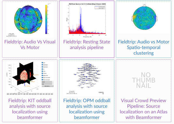
Gallery of published Jupyter notebooks covering preprocessing, time and frequency analysis, source localization, and quality control on both SQUID and OPM data.
Collaborative platform Researchers, students, and external collaborators contribute directly through pull requests. Review, discussion, and revision happen in the open.
Open source documentation All content is publicly hosted on Read the Docs and backed by a GitHub repository. Anyone can read, cite, or fork the material.
Reproducible pipelines Each notebook is executable end to end with versioned dependencies, so any analysis in a paper can be rerun from the raw data.
Section 25
How we work?
Automated Actions
Workflows under .github/workflows in the neurowaves lab documentation repository run on every push, every pull request, and on cron schedules. The human writes content and code; automation handles the rest.
BIDS dataset validation Every night at 20:00 UTC, datasets on Box are pulled and checked against the BIDS specification. Naming, metadata, and file structure issues are caught before analysis starts. bids-validation-deno.yml
Booking calendar sync Every 5 minutes, the Booked CTPSS scheduler is mirrored to Google Calendar from the MEG workstation, so lab users see one source of truth for reservations. sync-gcal.yml
Documentation PDF production Dedicated workflows build versioned PDFs from the RST sources: full lab documentation, MEG lab manual, CTP SOP and so on. report-generation-*.yml · 4 workflows
Read the Docs daily rebuild Every day at 10:00 UAE time the neurowaves Read the Docs project is retriggered so dashboards refresh with the latest empty room recordings and sensor diagnostics. readthedocs-daily-build-trigger.yml
Pipeline regression tests The MNE based pipelines are tested in a matrix on Ubuntu, macOS, and Windows with Python 3.11, so upstream library updates never silently break our analyses. test_pipelines.yml
Box dataset inventory and slide deploy On demand, the Box data lake is inspected and catalogued, and this CTP presentation itself is built and published with the NeuroWaves documentation. box-dataset-info.yml · deploy-docs-cloudflare.yml
Why it matters · automation enforces BIDS compliance, keeps bookings and dashboards always current and catches pipeline regressions early, so researcher effort stays focused on science.
Section 26
Next steps
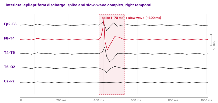
Epilepsy Research with Cleveland Clinic
NYUAD and Cleveland Clinic Abu Dhabi · multi modal neuroimaging of medically refractory focal epilepsy
Illustrative interictal spike, right temporal focus. Patients referred from CCAD for multimodal imaging at NYUAD.
Study
PI Prof. David Melcher (NYUAD)
Duration Oct 2025 to Oct 2028
IRB Abu Dhabi DOH approved
Imaging MEG, EEG, structural MRI, fMRI, simultaneous EEG plus fMRI
Spatiotemporal signatures of ictal and interictal activity, if they occurred
Research tasks before and after SEEG electrodes
Section 27
Next steps
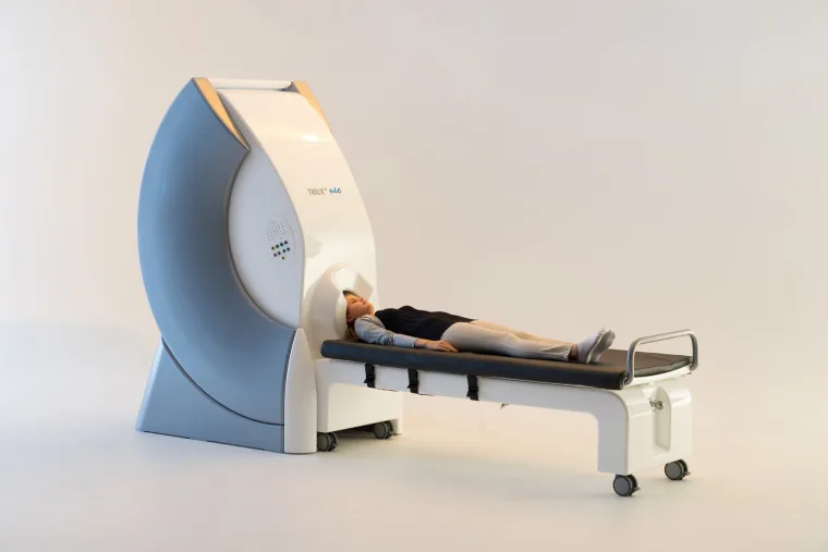
Procurement of a new SQUID system
Adding a MEGIN TRIUX neo clinical grade SQUID MEG at NYUAD.
Simultaneous EEG and MEG Integrated 128 channel EEG recorded in the same helmet as the 306 channel MEG array, so evoked activity is captured by both modalities at once.
Clinically validated FDA cleared and CE marked for pre surgical epilepsy mapping, enabling NYUAD to support the Cleveland Clinic Abu Dhabi clinical pipeline.
The Slidev source, document generator, custom Vue components, and presentation
assets live in the repository under
docs/source/7-meg-class-talks-demos/talks/ctp-presentation/.

 SQUID-MEG
 OPM-MEG